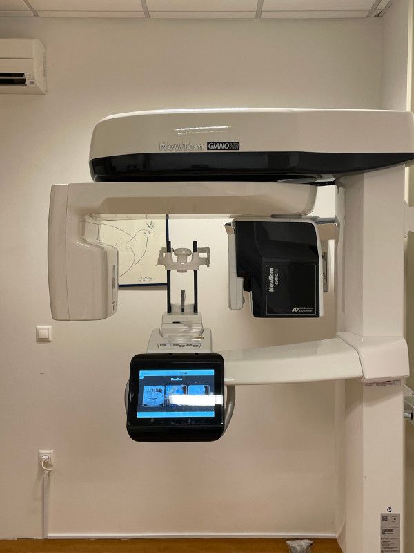
Dental Imaging
Chios MRI Diagnostic Imaging Center provides for all digital imaging examinations in one dimension (panoramic and cephalometric radiographs) and images in three dimensions (Dental CT).
The modern digital panoramic and cephalometric radiography system ORTHOPHOS 3C from SIRONA, provides the most reliable information in dental imaging in one dimension.

The examination is also provided in a digital format (CD or email), in order to process and make direct measurements on the dentist's computer.
The panoramic radiograph shows in a single image, teeth and bone structures of the maxilla & mandible and gives important information about the nasal area, the maxillary, the position of the teeth and gums and bone defects. The examination is also recommended for designing prostheses, exports and implants.
Η συγκεκριμένη οδοντιατρική απεικόνιση μπορεί επίσης να αποκαλύψει:
-
advanced periodontal disease
-
dental cyst
-
tumors and oral cancer
-
Impacted teeth
-
temporomandibular disorder
-
sinusitis
With the Multislice CT scanner SOMATOM SPIRIT and the appropriate software, a multilevel examination of teeth maxilla and mandible is available.
Computed tomography for dental purposes is recommended for the evaluation of patients with dental implants, tumors, cysts, inflammations, fistulas, silicone implants, fractures and surgeries. It provides accurate information on the height and width of the upper and lower jaw as well as positions of vital importance structures, such as the mandibular canal, the mandibular foramen, the foramen, the incisive canal and maxillary sinus. Moreover, detailed imformation emerges about the internal anatomy and the relationship between lesions, cementum bark and teeth roots. The images produced are of superb clarity because the technical errors (artifacts) from the fillings are eliminated because the final image in the coronal plane proginates from annealing images on a transverse plane.
Additionally, the system of Magnetic Resonance Imaging MAGNETOM C! can measure the temporomandibular joint position in an open and closed mouth, for examining arthritis lesions and mobility of the joint.


